Fig. 2.
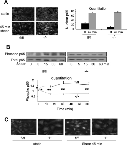
FAK regulation of NF-κB. Control FAKfl/fl (fl/fl) cells and FAK−/− cells were exposed to 24 dyn/cm2 laminar shear stress (LSS) for 45 min. A: cells were fixed and stained for p65, and >100 cells per condition in each experiment were scored for nuclear translocation. Scale bars = 50 μm. Values are means ± SE (n = 3). B: cells were exposed to flow for the indicated times and p65 Ser536 phosphorylation was analyzed. Results were quantified and normalized to total p65 levels. Values are means ± SE (n = 6). *P < 0.05, **P < 0.01. C: cells with or without 45 min of flow were fixed and stained for phospho-Ser536 p65. Images are representative of 3 independent experiments.

