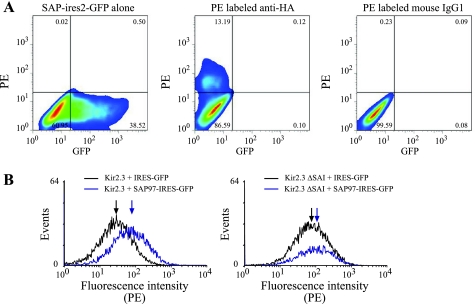
Fig. 6.
Introduction of SAP97 in Kir2.3-expressing HEK cells increases channel protein abundance and cell surface expression. A: HEK cells were transfected with SAP97-IRES2-GFP or hemagglutinin antigen (HA)-tagged Kir2.3. Left: cells expressing SAP97-IRES2-GFP. Cells transfected with HA-tagged Kir2.3 were stained with either a PE-conjugated anti-HA antibody (middle) or a PE-conjugated isotype control antibody (right). The percentages of cells in each of the four quadrant gates is indicated. B: cell surface expression of Kir2.3 in HEK cells transiently transfected with HA-tagged Kir2.3 or HA-tagged Kir2.3ΔSAI and pIRES-GFP (black) or pSAP97-IRES-GFP (blue). Surface expression of Kir2.3 was detected using an PE-conjugated anti-HA antibody. Gating parameters were adjusted to display the surface expression of Kir2.3 in the GFP positive cells. The median fluorescence intensity for each population is indicated by an arrow. The data shown are representative of two separate experiments. Removal of the COOH-terminal PDZ-binding motif (HA-tagged Kir2.3ΔSAI) attenuated the SAP97-induced increase in cell surface expression in transiently transfected cells.

