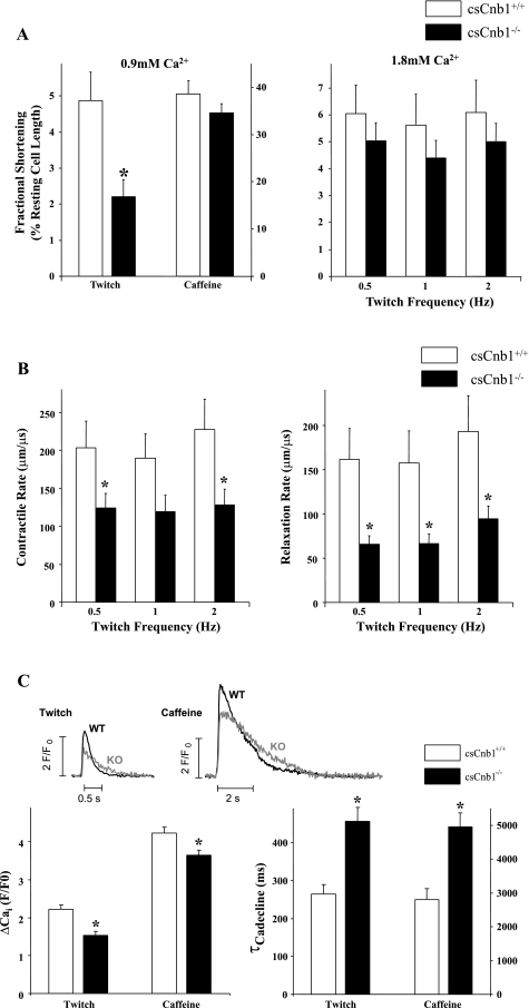
Fig. 7.
Altered contractile parameters of isolated adult cardiac myocytes from csCnb1−/− mice. A: mean contraction amplitudes as a percentage of total cell length of cardiac myocytes (n = 13 cells per group) isolated from csCnb1+/+ and csCnb1−/− ventricles measured at 1 Hz and 0.9 mM Ca2+ (left) or 0.5–2 Hz and 1.8 mM Ca2+ (right). B: mean rates of contraction (left) and relaxation (right) of cardiac myocytes maintained in 1.8 mM Ca2+ and stimulated at 0.5–2 Hz. C: Ca2+ transients induced by electrical stimulation (0.5 Hz) or rapid exposure to 10 mM caffeine (0.9 mM Ca2+ concentration). Amplitudes (left) and time constants (τ) of intracellular Ca2+ (Cai) concentration decline (right) are shown. *P < 0.05 compared with controls at the same contraction frequency. WT, wild type; KO, knockout; F/F0, ratio of background fluorescence to diastolic fluorescence under control conditions.

