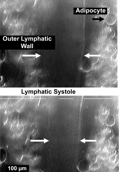
Fig. 1.
A video image of an intact, in vivo lymphatic in diastole (top) and at full systole (bottom). The mesenteric lymphatic vessels associated with major mesenteric arteries are virtually surrounded by adipocytes, and the exact inner diameter locations are difficult to resolve. The white broken line along the top of each image was generated by the Living Systems Dimension Analyzer and is part of an automated system to track changes in dimensions, in this case, the edges of adipocytes adhered to the lymphatic. Normally, the 2 longer lines would terminate before crossing over the outer walls of the lymphatic. The limited contrast of the images, despite oblique lighting and modification of the image with the camera controls, made simultaneous measurements of both wall locations and thereby diameter difficult. However, 1 of the 2 measurement windows did consistently track motion to allow the start of systole to be accurately determined.

