
Figure 1.
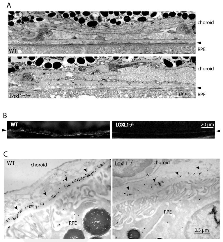
(A) Transmission electron micrographs showing the elastic lamina of Bruch's membrane in WT and LOXL1 mutant mice (at 3 months of age). Note the continuous elastic lamina in the WT in contrast to the many discontinuities (gaps) seen in the mutant. (B) Immunofluorescence staining for elastin in Bruch's membrane. WT (left) and LOXL1 mutant (right) mice show different amounts of elastin staining (arrowheads) in the elastic lamina of Bruch's membrane. The samples were lightly fixed and extracted with a denaturant (urea). In the WT, elastin immunostaining appears as a bright line that is continuous along Bruch's membrane. Elastin staining appears much weaker in the mutant. (C) Immunogold labeling of elastin in Bruch's membrane. Representative images of WT and LOXL1-deficient mice are shown. Elastin labeling (arrowheads) in the mutant mouse is scanty and fragmented, whereas that in the WT is more continuous and of a higher density.