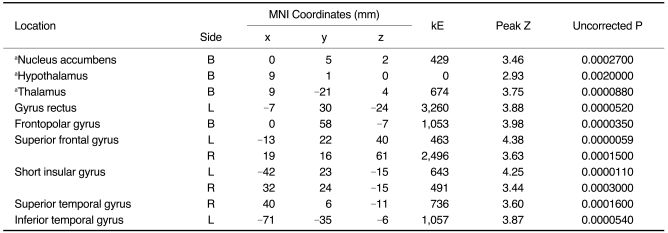
Table 2.
Brain Regions Showing Significant Decrease in Gray Matter Concentration by Voxel-Based Morphometry in Narcoleptics with Cataplexy
Note.-MNI = Montreal Neurological Institute, B = bilateral, L = left, R = right. Height threshold, uncorrected p < 0.001. Extent threshold kE > 100
aSignificant at false discovery rate p < 0.05 by small volume correction using sphere of radius 30 mm located at center point (x, y, z: 0, 0, 0).

