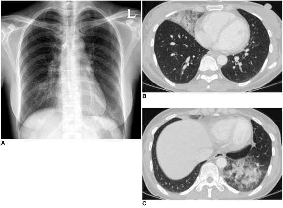
Fig. 2.
42-year-old woman diagnosed as novel influenza A (H1N1) pneumonia with secondary pneumococcal pneumonia.
A. Initial chest radiograph shows ill-defined infiltrates in both lower lung zones.
B, C. Chest CT scans show lobar-distributed ill-defined consolidation and peripheral ground-glass opacities in right middle lobe and left lower lobe.

