Figure 3.
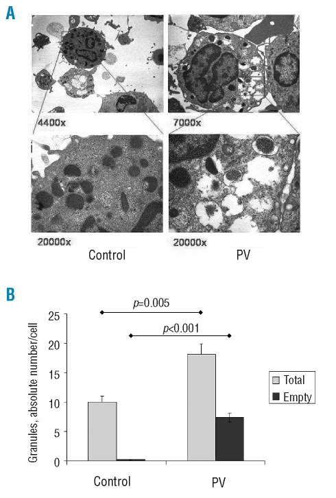
(Panel A) Representative transmission electron microscopy analysis of circulating basophils in a control subject (images on the left) and a PV patient (on the right; V617F allele burden =70%). Thin (upper panels) and ultrathin (lower panels) sections were observed under vacuum with an EM 109 Zeiss microscope equipped with built-in electromagnetic objective lenses and camera (Oberkochen, Germany). Photographs were taken with Kodak Technical Pan film (Kodak, Rochester, NY, USA), developed with Kodak D 19 1+4 automatic developer and scanned with an EPSON Perfection 3200 photoscanner (Seiko EPSON, Nagano-ken, Japan). Original magnification was 4,400× and 7,000× for the upper left and right panel, respectively, and 20,000× for the lower panels. (Panel B) The absolute numbers of granules contained in basophils from PV patients (n=5) and healthy subjects (n=4) (gray columns) after enumerating at least ten basophils/subject; the numbers of those granules devoid of their electron-dense content (empty granules) are also presented (black columns). Statistically significant differences are reported in the plot.

