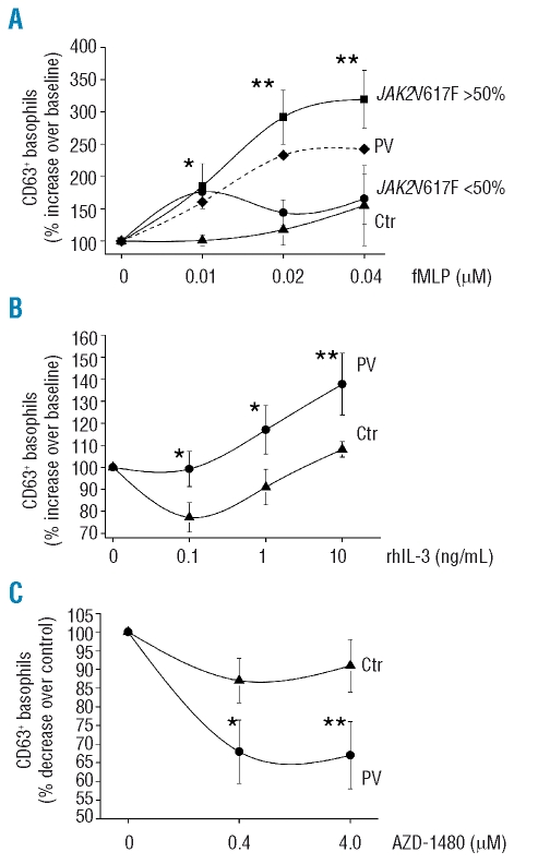
Figure 4.
(Panel A) Expression of the activation marker CD63 in peripheral blood cells after being incubated ex vivo with increasing amounts of fMLP peptide (0 to 0.04 μM) in the presence of an optimal amount of rhIL-3 (10 ng/mL). Results are expressed as per cent increase of CD63+ basophils over unstimulated cells. The mean (±SD) values measured in control subjects (n=5; triangles) and PV patients (n=10), either all together (dashed line; for clarity, SD is not presented) or divided according to their V617F allele burden [>50% (squares) or <50% (dots), n=5 each], is presented. (Panel B) Experiments as above were performed using increasing amounts of rhIL-3 in the presence of a fixed dose of fMLP peptide (0.02μM). Only PV patients with more than 50% mutated V617F allele were included in these experiments and compared to controls (n=5 each). Results are expressed as per cent increase of CD63+ basophils over cultures containing fMLP only. (Panel C) Peripheral blood cells from PV patients and control subjects (n=5 each) were pre-incubated with the specific JAK2 inhibitor AZD1480 at two different concentrations, and then challenged with fMLP peptide (0.04 μM) and IL-3 (10 ng/mL). The fraction of cells in the basophil gate expressing CD63 was measured by FACS; results are expressed as per cent decrease of CD63+ basophils in wells containing the drug compared to cells without inhibitor. Only PV patients with more than 50% mutated V617F allele were used in these experiments. *p<0.05; **p<0.01.

