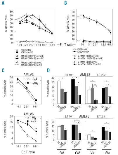
Figure 2.
(left). Cytotoxicity of NK cells against AML-LSCs and N-HSCs. (A-D) Single-KIR+ NK cells were used as effectors in chromium-release assays. (A) AML-CD34+CD38− LSCs and CD34+CD38+ blasts from patient AML#8 or control K562 cells were exposed to NK cells, matched (mNK, KIRe+ NK-1) or mismatched (mmNK, KIRa+ NK-1), at the indicated E:T ratios. (B) N-BM CD34+ and N-mPB CD34+CD38− HSCs from 2 healthy donors, or control K562 cells were exposed to NK cells, mNK (KIRe+ NK-2 for N-BM; KIRa+ NK-3 for N-mPB#1) or mmNK (KIRa+ NK-2 for N-BM; KIRb+ NK-3 for N-mPB), at the indicated E:T ratios. Availability of N-BM samples was limited and this did not allow selection for CD34+CD38− cells in numbers sufficient for the cytotoxicity assay. (C) AML-CD34+CD38− LSCs from patients AML#3 and AML#6, either non-treated (−VA) or incubated for two days with VA (+VA) were exposed to mmNK cells (KIRe+ NK-1) at the indicated E:T ratios. (D) AML-CD34+D38− LSCs from patients AML#3 and AML#6 were incubated for two days with medium alone (M; ○), or with VA (▪) and exposed to mmNK cells (as in C) which were blocked by preincubation with control mouse IgG ( ) or aNKG2D mAb (▥) for 1 h at 37°C, at 10:1 E:T ratio. All experiments were performed in triplicates; mean±SEM values are shown. *p<0.05. The NK cell lines are defined in Online Supplementary Table S1.

