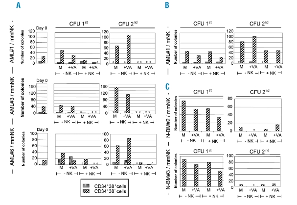
Figure 3.
Effect of NK cells on hematopoietic colony formation by AML-LSCs and N-BM HSCs. (A–C) Single KIR+ NK cells were used as effectors and their effect on growth of CFU from AML-LSCs and N-BM HSCs was tested in 1% methylcellulose cultures containing erythropoietin (3U/mL), interleukin (IL)-3 and -6, granulocyte- and granulocyte-macrophage-colony stimulating factor (20 ng/mL, each), stem cell factor and flt3 ligand (100 ng/mL, each). AML-CD34+CD38− LSCs (1×10⁵) and CD34+CD38+ blasts (1×10⁵) from 3 patients (AML#1, AML#3 and AML#6), or N-BM CD34+CD38− HSCs (1×10³) from 2 healthy donors (N-BM#2 and #3) were incubated for two days with medium alone (M) or VA (+VA) and plated in methylcellulose directly (−NK) or after exposure to NK cells (+NK), either matched (mNK) or mismatched (mmNK), for 4 h at E:T ratio of 5:1. (A) CFU numbers by AML cells prior to incubation (Day 0). A–C Numbers of CFU, primary 1st or secondary 2nd. (A) mmNK were KIRe+ NK-1. (B) mNK were KIRa+ NK-1. In C, mmNK were KIRa+ NK-1. The NK cell lines are defined in Online Supplementary Table S1. Results are shown as mean of duplicate analyses. Cultures in which no CFU-derived colonies were seen are indicated with “0”.

