Figure 5.
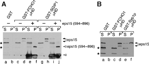
μHDs of FCHO1 and Syp1 bind eps15. (A) Approximately 200 μg of GST (lanes a, b), GST-FCHO1 μHD (lanes c–f) or GST-SGIP1 μHD (lanes g–j) immobilized on beads was incubated with clarified rat brain cytosol in the absence or presence (lanes e, f, i, j) of 25 μM eps15 (594–896) polypeptide as indicated. Aliquots of each supernatant (S) and washed pellet (P) were resolved by SDS–PAGE, transferred to nitrocellulose, and probed with anti-eps15 antibodies; only the relevant portions are shown. The eps15 competitor peptide and a degradation product (open arrowheads) are shown. Two non-specific bands detected by the antibody are indicated (asterisks). (B) Approximately 200 μg of GST (lanes a, b) GST-FCHO1 μHD (lanes c–d) or GST-Syp1 μHD (lanes e–f) immobilized on beads was incubated with clarified rat brain cytosol and analysed similarly.

