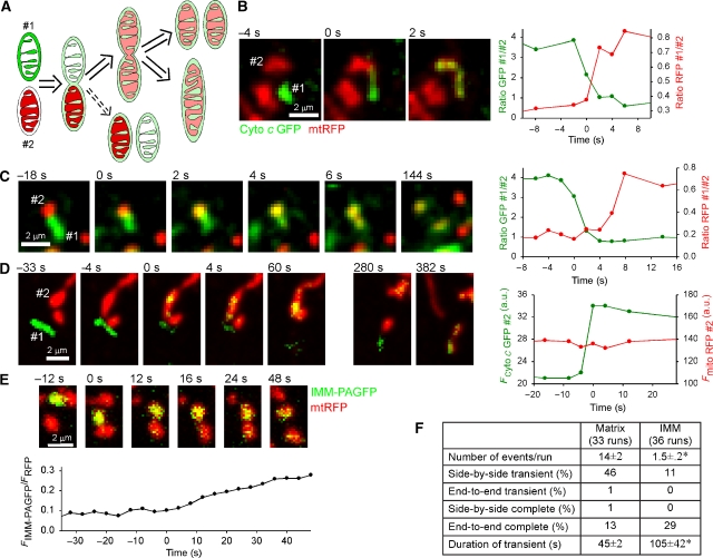
Figure 2.
Sequential and separable fusions of OMM and IMM observed during transient and complete fusion events. (A) Scheme showing the possible paths of transient/complete fusion observed by PEG fusion of cells expressing IMS-targeted cyto c GFP with mtRFP-expressing cells. Fusion of the OMM results in diffusion of the cyto c GFP into the ‘red' mitochondrion usually followed by IMM fusion and diffusion of the mtRFP into the ‘green' mitochondrion. In rare cases, the OMM fusion is reversed without IMM fusion having occurred (dashed arrow). (B) Close temporal coupling of OMM and IMM mergers observed with cyto c GFP and mtRFP as described above. The separation of events is not visible in the images, although the graph reveals an ∼1-s lag between exchange of IMS and matrix contents. (C) Clearly separated OMM and IMM fusions during transient fusion. Mitochondrion #2 becomes yellow from intake of cyto c GFP ∼6 s before mtRFP enters #1. (D) OMM fusion can be completely uncoupled from IMM fusion: an example of the rare OMM-only transient fusion. Mitochondrion #1 donates cyto c GFP to #2 and re-separates (60 s) without having merged matrices. Minutes later #1 undergoes a matrix fusion with another mitochondrion (382 s). (E) Slow and partial mixing of integral IMM proteins during transient fusion after photoactivation in H9c2 cell expressing both IMM-PAGFP and mtRFP. (F) Comparison of the transfer of soluble matrix and integral IMM proteins during transient and complete fusions in cells expressing mtPAGFP/mtRFP or IMM-PAGFP/mtRFP (mean±s.e.m.; *P⩽0.001).

