
Figure 5.
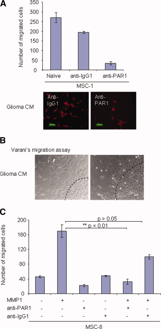
Functional MMP1/PAR1 axis mediates MSC migration. (A): Effect of anti-PAR1 blocking antibody on the migration of MSC-1 was examined using a modified Boyden chamber assay. Bar graph represents number of migrated cells. Data shown are averages of triplicates ± SEM, experiment was repeated independently three times. Photomicrograph showed the representative images of PI+ migrated cells at original magnification ×200. (B): Varani migration assay was performed to confirm the effect of PAR1 on MSC migration. Photomicrograph showed migration of MMP1-RNAi-transfected MSCs. Representative images from two independent experiments were shown. Images were shown as original magnification x100. Slides were visualized using wide-field microscopy with an inverted microscope (TE300; Nikon), and images were acquired on a CCD color digital camera (DXM1200F; Nikon) using image acquisition software, ACT-1 v2.7 (Nikon). (C): Effect of MMP1/PAR1 interaction on the migration ability of MSC-8 treated with the various proteins was examined using a modified Boyden chamber assay. Bar graph represents number of migrated cells. Data shown are averages of triplicates ± SEM, experiment was performed independently twice. Abbreviations: CM, conditioned medium; MSCs, mesenchymal stem cells; PAR1, protease-activated receptor one.