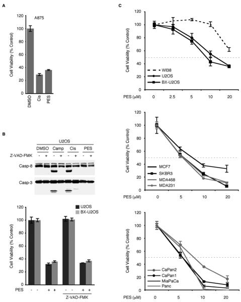
Figure 3. PES Reduces Viability of Tumor Cells and Induces Cytoplasmic Vacuolization.
(A) MTT assays of A875 cells treated with DMSO, 20μM PES, or 50μM cisplatin for 24 h. Each graphical representation indicates the mean ± SD of at least three independent cultures relative to control (DMSO-treated) cells.
(B) (Top) WCE were prepared from U2OS osteosarcoma cells that were either untreated or pretreated with Z-VAD-FMK (20μM) for 1 h, followed by the addition of 5μM camptothecin (Camp), 16μg/ml Cisplatin (Cis), or 20μM PES for 6 h. Note evidence of caspase cleavage in cisplatin- or camptothecin-treated cells, but not in the presence of PES or Z-VAD-FMK. (Bottom) The indicated cell lines were either untreated or pretreated with Z-VAD-FMK (20μM) for 1 h, followed by the addition of DMSO or 20μM PES for 24 h. Cell viability was determined by MTT assays. Results shown are the mean of at least three independent experiments.
(C) The indicated cell lines were treated with the indicated concentrations of PES for either 24 h (top) or for 48 h (middle and bottom). Representative MTT assays indicate cell viability in human cell lines, including non-transformed human WI38 fibroblasts, as well as several tumor cell lines with wt p53 (U2OS, BX-U2OS, MCF7, CaPan2), or with mutant/deleted p53 (SKBR3, MDA468, MDA231, CaPan1, MiaPaCa, and Panc1). Four independent cultures were assayed for each treatment and DMSO treated cells were used as internal controls. Values shown are normalized to the viability of the control (DMSO-treated) cells. Error bars represent standard deviation (SD).

