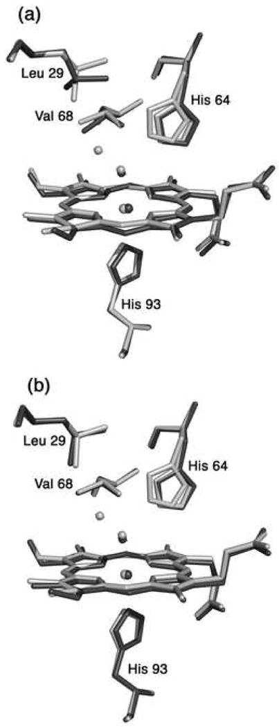
Fig. 3.

A superposition of the heme environments in the crystal structures of hh MnIIIMb(H2O) (this work, shown in light grey) and (a) natural hh FeIIIMb(H2O) (pdb code 1YMB [80], shown in dark grey), and (b) recombinant hh FeIIIMb(H2O) (pdb code 1WLA [79], shown in dark grey), using a global Cα structural alignment. The major conformation of His64 in hh MnIIIMb(H2O) is similar to the conformation in FeIIIMb(H2O). The Leu29 and Val68 conformations of the Mn- and Fe-proteins overlay better in the structures shown in Fig. 3b.
