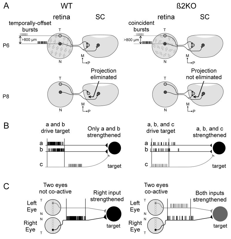
Figure 6.
Models of how retinal waves mediate the refinement of visual system projections. (A) Synapse elimination: In WT mice, the directional bias of retinal waves along the NT axis during the first post-natal week helps refine projections along this axis by ensuring that RGCs separated by 800+ μm are more likely to fire temporally-offset bursts (top left, wave direction designated by arrows in retina; see Figure 4). This helps eliminate topographically incorrect projections by the second week (bottom left). Because the directional bias of waves is lost in β2KOs, this decreases the likelihood that RGCs separated by 800+ μm will fire temporally-offset bursts (top right). Topographically incorrect synapses are not eliminated and axonal projections fail to refine along the NT axis (bottom right). (B) Synapse strengthening: In WT retinal waves, short ISIs and strong local correlations within each eye ensure that only neighboring RGCs (a and b) drive post-synaptic bursting in target neurons, stabilizing these synapses but not ones from distant RGCs (c, left). In β2KOs, longer correlations allow distant RGCs (a,b, and c) to drive coincident post-synaptic spiking, stabilizing misdirected axonal projections (right). (C) Eye-specific segregation: In WT retinas, the lack of RGC activity during inter-wave intervals ensures that retinotopically matched regions of the two eyes are not co-active. This helps strengthen projections from one eye, thereby segregating inputs from the same eye (left). In β2KOs, inter-wave activity is increased leading to an increased probability of co-active inputs. This strengthens inputs from both eyes and segregation fails to occur normally (right).

