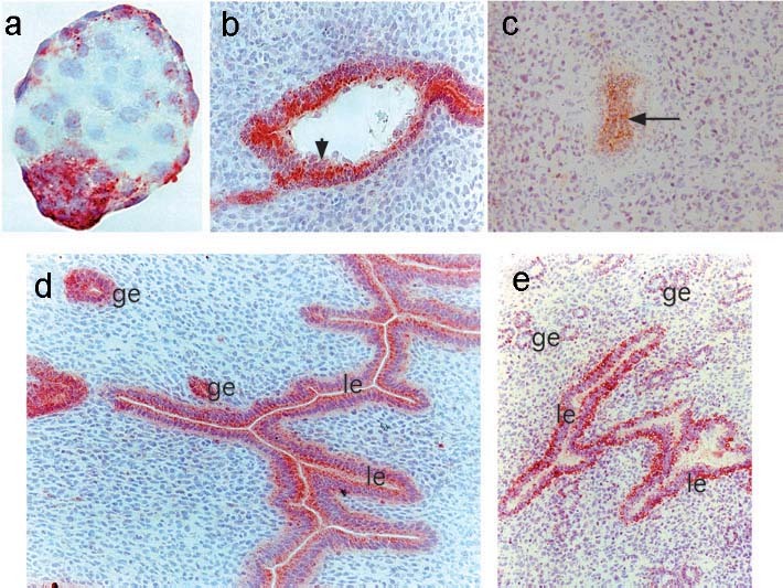
Figure 2.

Expression of bystin protein in mouse embryos before, during and after implantation. (a) Immunohistochemistry for bystin in a blastocyst. Bystin protein was found in a blastocyst but not detected in a fertilized mouse egg or embryos earlier than the blastocyst stage [30]. (b) Bystin protein was barely detected in trophectoderm cells (arrowhead) during implantation. (c) After implantation, bystin protein was found in the epiblast including embryonic stem cells (arrow). (d) In the pregnant mouse uterus or in the presence of blastocysts, bystin expression is seen on the apical side of luminal epithelia (le), whereas it is distributed evenly in glandular epithelia (ge). (e) Immunohistochemistry of the uterus of a non-pregnant female mouse. Note that bystin proteins were found at the abluminal side of endometrial luminal epithelia (le) and glandular epithelia (ge). Tissue sections were reacted with rabbit anti-bystin antibody [30], followed by biotinylated antirabbit IgG antibody and peroxidase avidin. The peroxidase substrate 3-amino-9-ethyl carbazole was used to detect immunostaining. Hematoxylin was used for counter-staining.
