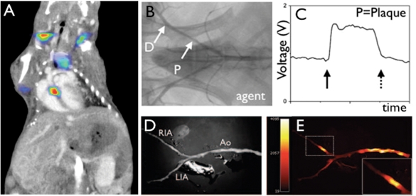
Figure 1.
Fluorescence imaging of protease activity. (A) In vivo fluorescence molecular tomography (FMT) of an atherosclerotic mouse after injection of a cathepsin-targeted protease reporter. Hybrid FMT–CT imaging shows activation of the sensor in the aortic root, a site of strong inflammatory atherosclerosis in this model.76 (B–E) Catheter-based sensing of fluorescence protease reporters in a rabbit model of arterial injury. The balloon injury was applied in the iliac artery (B). During pull back of a fluorescence sensing catheter, high signal was observed (C), corroborated by ex vivo fluorescence reflectance imaging (D, E).13

