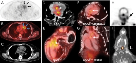
Figure 2.
Nuclear imaging of plaque biology. (A–C) Clinical 18FDG PET–CT imaging in a patient with coronary artery disease shows uptake of the tracer in a stenotic segment (confirmed by angiography).19 (D–G) In vivo PET–CT imaging with the VCAM-1 targeted tracer 18F-4V in atherosclerotic mice with and without statin treatment. (D, F) Short-axis view of aortic root, (E and G) long-axis view. Statin therapy reduced VCAM-1 expression and consecutively PET signal. (H) SPECT imaging of apoptosis in the carotid artery of a TIA patient.27 (I) In vivo MMP-targeted microSPECT-CT 3 weeks following carotid injury in the apoE−/− mouse. Arrows point to the injured left (L) and non-injured right (R) carotid arteries.28

