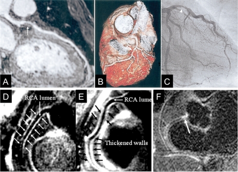
Figure 3.
Visualization of a coronary atherosclerosis by MRI. (A) Curved multiplanar reconstruction of a whole-heart coronary MRA data acquisition shows a stenosis in the left anterior descending (LAD) artery (arrow). (B) Volume-rendering method in the same patient demonstrates three-dimensional view of the LAD with stenosis (white arrows). (C) X-ray coronary angiography confirms the stenosis of the proximal LAD (arrowhead),38 reprinted with permission from Elsevier. (D and E) Coronary vessel wall images of the proximal RCA in two other subjects without coronary luminal stenoses; (D) a 58-year-old man with long-standing type 1 diabetes and normoalbuminuria and (E) a 44-year-old man with long-standing type 1 diabetes and diabetic nephropathy. In (D), there is no evidence of atherosclerotic plaque. However, an increased atherosclerotic plaque burden is seen in (E). The anterior and posterior RCA walls are indicated by arrows44 reprinted with permission from Wolters Kluwer Health. (F) Delayed focal gadolinium late enhancement of the proximal RCA (arrow) is shown in another patient with X-ray defined coronary artery disease in the same location,48 reprinted by permission of the European Society of Cardiology.

