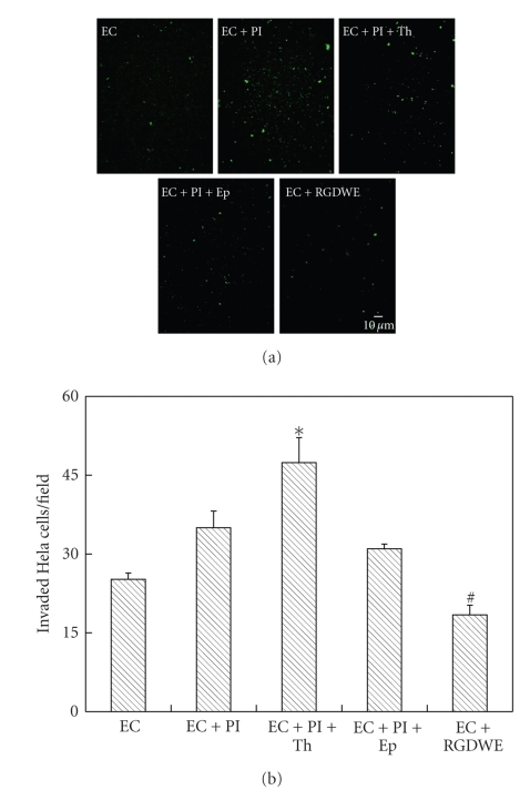
Figure 6.
Transmembrane invasion of HeLa cells through PET membrane precoated with gelatin and a monolayer of HUVECs cultured in the outer side of PET membrane. The numbers of invaded HeLa cells (labelled into green) were quantified by cell counting from more than five micrographic fields using magnification of 10×. EC: HUVECs cultured on the outer side of PET membrane. EC+Pl: in the presence of HUVECs monolayer and platelet; EC+Pl+Th: in the presence of HUVECs monolayer and thrombin-activated platelets; EC+Pl+Ep: in the presence of HUVECs monolayer and thrombin-activated platelets treated with eptifibatide; EC+RGDWE: in the presence of HUVECs monolayer and HeLa cells with RGDWE peptides blocked αvβ3 integrin. *P < .05 (EC+Pl+Th) versus (EC+Pl) or (EC+Pl+Ep); #P < .05 (EC+RGDWE) versus EC.

