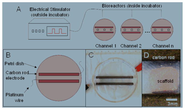
Fig. 1.
Experimental setup for applying pulsatile electric field stimuli to cardiac cells. (A) Overview of experimental setup. An electrical stimulator generates the pulses which are transmitted to bioreactors located inside an incubator maintained at 37° C. (B) Schematic diagram of the electrical stimulation chamber (modified 60 mm Petri dish with carbon rod electrodes). 3-D scaffolds may be placed in between the electrodes. (C) Photograph of assembled electrical stimulation chamber. (D) Close up view of scaffold positioned between electrodes.

