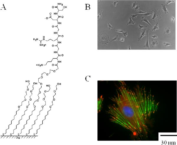
Figure 2.
(a) Structure of a monolayer that presents the peptide Arg–Gly–Asp mixed with tri(ethylene glycol) groups. (b) An optical micrograph that shows 3T3 fibroblasts attached to a monolayer wherein 0.5% of the alkanethiolates present the peptide ligand. (c) A fluorescent micrograph of a cell that was adherent on these monolayers for 12 h, fixed and stained with phalloidin-rhodamine (green) to reveal the actin cytoskeleton and with anti-vinculin (red) to reveal focal adhesion complexes. The cells assembled stress fibers that were indistinguishable from those found in cells adherent on fibronectin.

