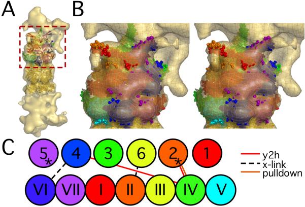
Figure 3.
AAA-ATPase-CP model. A: The atomic model of the AAA-ATPase-CP sub-complex fitted into the cryo-EM map of the 26S proteasome. The AAA-ATPase model is placed on the top. The six AAA-ATPase subunits and the adjacent CP α-subunits are colored as indicated in panel C, the remaining CP subunits in gold. The AAA-ATPase quaternary structure corresponds to Fig. 2C. B: Zoomed-in stereo view of the rectangular area in A. C: Schematic view of the topology of the CP-AAA-ATPase model and the CP-RP interactions (Table 1). The AAA-ATPase subunits are displayed in Arabic numbers, whereas Roman numbers indicate the CP α-subunits. The asterisks mark the AAA-ATPase subunits that have been shown to induce gate opening of the CP (Rpt2 and Rpt5).

