
Fig. 2.
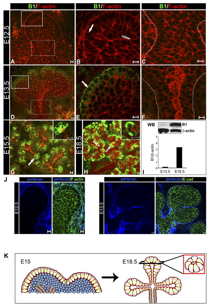
The outer cell layer expresses acinar marker, B1, early in SMG bud development. SMGs representing E12.5, E13.5, E15.5, and E18.5 stages of embryonic development were immunostained with anti-B1 antibodies (green), counterstained with rhodamine-phalloidin (red) and examined by confocal microscopy. A–C: Cells in the E12.5 SMG were not reactive with B1 antibodies. D–F: Immunolocalization of B1 in the E13.5 SMG was restricted to the peripheral cell layer (D, white solid box; E, block arrow) while the stalk region was negative (D, white dashed box; F, region within dashed lines). G: Immunolocalization of B1 in the E15.5 SMGs was maintained in the outer layer of the developing buds (block arrow; inset: 63×). H: At E18.5 acinar cells had prominent B1 expression (block arrow; inset: 63×) while ducts remained negative (unfilled arrow). I: Expression of B1 was detected early in morphogenesis and increased in the cytodifferentiated SMG. Glands were extracted with Triton extraction buffer and analyzed by Western blot (WB) using anti-B1 and anti-β-actin antibodies. Bar graph represents B1 normalized to β-actin; shown is one of two independent experiments. J: Perlecan staining of the basement membrane of the early SMGs. E12.5 and E13.5 SMGs were immunostained for perlecan (blue) and either E-cadherin (green) or F-actin (green). Perlecan (blue) stains the basement membrane, which is in direct contact with the outer layer of the SMG epithelium. Shown are middle 1-μm optical sections. Scale bar = 10 μm. K: A model showing how acinar cell precursors in the outer layer (white cells) develop into acini at the completion of SMG embryonic morphogenesis. Scale bar = 10 μm.