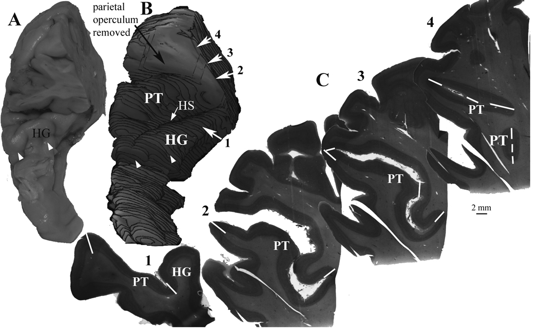
Figure 1.
An example of the methods used to measure the gray matter volume of the planum temporale (PT). A) The full extent of the PT and Heschl’s gyrus was dissected from each hemisphere. The parietal operculum was also included to prevent loss of any part of the caudal end of the lateral sulcus. The perspective in this image is from the anterior and dorsal view. B) For each hemisphere, a 3-dimensional reconstruction was made from video images of the block face during sectioning. Reconstructions were made from the every 12th section, using the same series of sections used for Nissl staining. The parietal operculum was removed from the reconstruction in order to view the PT surface. Heschl’s sulcus (HS) separates Heschl’s gyrus from the PT. The rostral end of Heschl’s gyrus is typically seen as an obvious end of the gyrus (white arrowheads in A and B). C) Nissl stained sections were digitally imaged, and the volume of cortex extending from Heschl’s gyrus (HG), or from the fundus of the lateral sulcus, to the lateral crest of the lateral sulcus was included as the PT. The white lines (solid and dashed) show the borders of the PT that were used to delimit the PT from adjacent structures, as described in the Methods section. Care was taken to include all cortex extending to the terminal end of the lateral sulcus. In this example, the numbered sections are the same as the corresponding sections in B.

