
Figure 3.
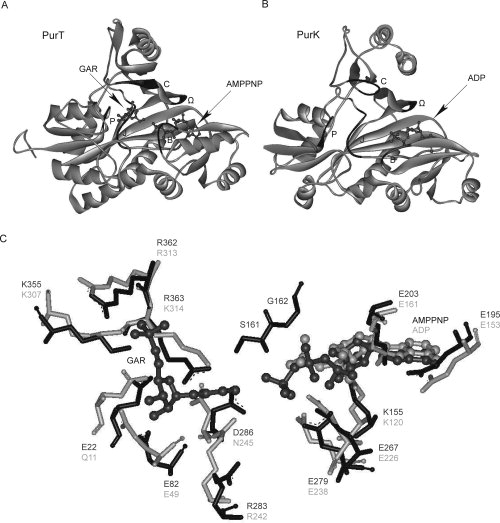
Overall architecture of the active sites in PurT (A), PurK (B), and residues within the PurT and PurK active sites (C). In (A) and (B), the B-loop, the Ω-loop, the P-loop, the J-loop, and the C-loop, are shown in black, with the backbones in gray. The GAR, AMPPNP, and ADP molecules are shown in ball-and-stick representation, and colored in dark gray. In (C), the residues involved in substrate binding and catalysis in the PurT (black) and PurK (gray) active sites are shown. Residues are numbered based on the Escherichia coli enzymes. Superpositioning of the PurT and PurK structures was done manually using Weblab Viewer Pro 3.7 (Molecular Simulations Inc.).