
Figure 2.
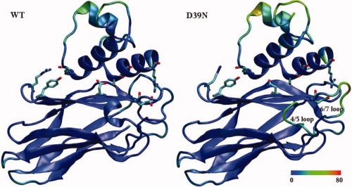
Time-averaged structures of the WT Cohesin-Dockerin complex and the D39N mutant resulting from MD simulations. Colors are assigned by B-factor from low-mobility (blue) to high-mobility (red), with the color scale in Å shown at the bottom. Carton representations are shown for both the WT and D39N mutant. Key residues are highlighted in licorice mode and colored by atom name.