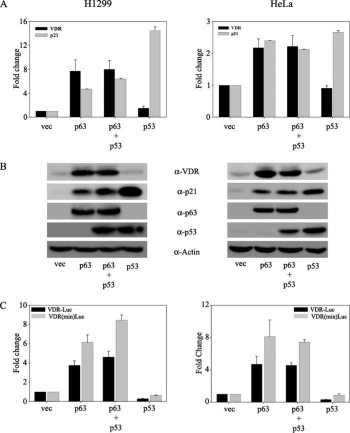
FIGURE 1. p53 does not affect the p63-mediated induction of VDR.
H1299 and HeLa cells were transfected with empty vector (vec), p63, p53, or p63 and p53 together as indicated. A, at 24 h post-transfection, total RNA was harvested and subjected to TaqMan reverse transcription-PCR. Glyceraldehyde-3-phosphate dehydrogenase transcript levels were used as normalization for the VDR mRNA levels. The y axis represents -fold change in VDR and p21 transcript levels relative to empty vector-transfected cells. B, whole cell extracts of transfected cells were subjected to immunoblot analysis using anti-VDR, anti-p21, anti-p53, and anti-p63 antibodies. Immunoblot analysis for β-actin served as the loading control. C, H1299 and HeLa cells were co-transfected with either full-length or the minimal VDR promoter construct along with either empty vector or expression plasmids encoding p63 alone or with p53 or p53 alone as indicated. A constant amount of expression plasmid encoding Renilla luciferase was included in all transfections to normalize for transfection efficiency. At 24 h post-transfection, cells were harvested in passive lysis buffer (PLB) and subjected to dual luciferase assays as per the manufacturer's protocol. RLU/R-Luc ratios were calculated to normalize for transfection efficiency. The y axis represents -fold change relative to empty vector control.

