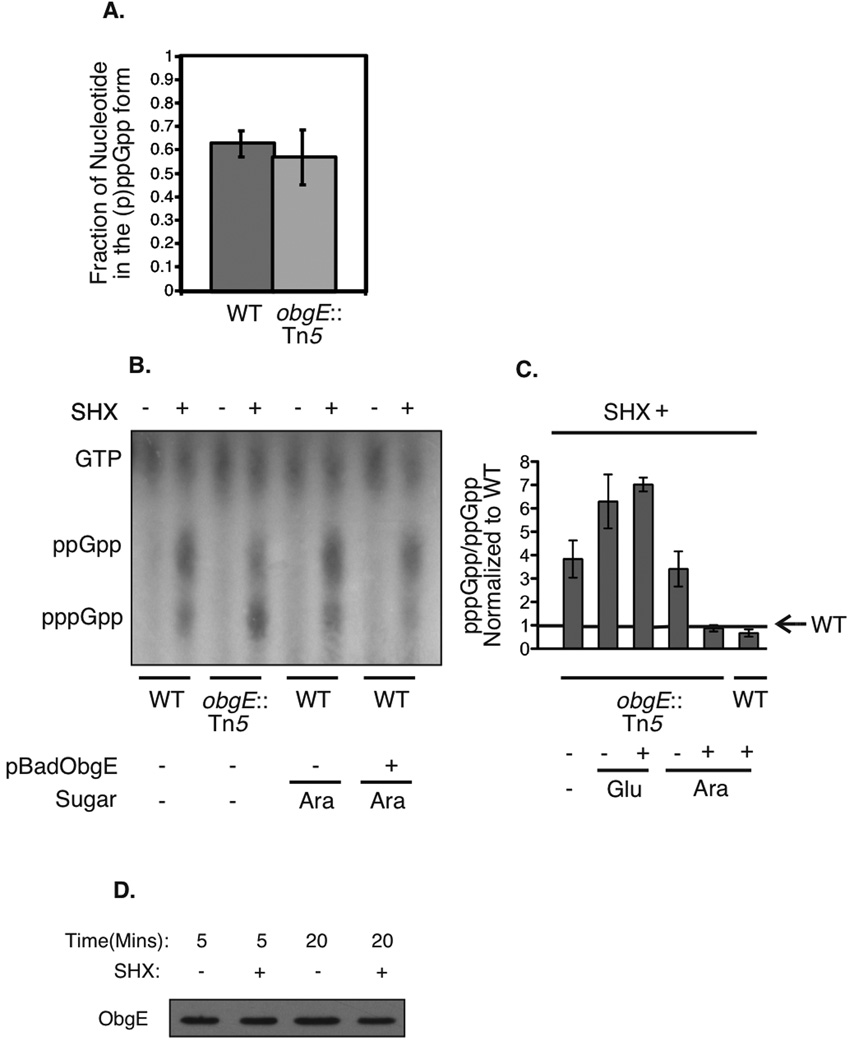
Figure 2.
Levels of (p)ppGpp in cells treated with serine hydroxamate. A) Fraction of total guanine nucleotides in the (p)ppGpp form for wild-type and obgE::Tn5 mutant cells, averaged from three independent experiments. Error bars represent the standard deviations of the means. B) Representative thin layer chromatography plate for in vivo ppGpp and pppGpp detection, with and without serine hydroxamate treatment. The samples shown (from left to right) are: wild type (MG1655), obgE::Tn5 (STL7742), wild type (MG1655) grown in the presence of arabinose, wild-type expressing pBADObgE+ (STL7679) grown in the presence of arabinose. C) Cumulative data averaged over three experiments each showing the ratio of pppGpp/ppGpp following serine hydroxamate treatment, normalized to wild type (MG1655). Error bars represent standard deviations of the means. The samples shown (from left to right) are: obgE::Tn5 (STL7742), obgE::Tn5 (STL7742) grown in the presence of glucose, pBADObgE+ in an obgE::Tn5 background (STL13897) grown in the presence of glucose (plasmid expression off), obgE::Tn5 (STL7742) grown in the presence of arabinose, pBADObgE+ in an obgE::Tn5 background (STL13897) grown in the presence of arabinose (plasmid expression on), and pBADObgE+ in a wild type background (STL7679) grown in arabinose (plasmid expression on). D) Levels of ObgE protein before and after serine hydroxamate treatment. Samples were taken at time-points following the addition of serine hydroxamate and ObgE was detected via western blot using polyclonal anti-ObgE antibody.

