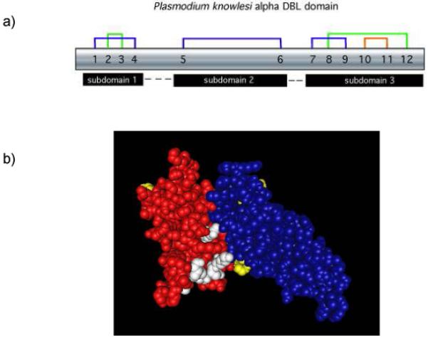
Figure 1. Subdomain structure of the P. knowlesi DBPα DBL domain is defined by disulfide bonding.

(A) Disulfide bonds, numbered sequentially 1-12, create three subdomains within the DBL ligand domain. (B) The subdomains highlighted by separate colors are connected by flexible linkers and weak hydrophobic forces maintain the subdomain configuration. In this model subdomain 1 is yellow, subdomain 2 is red, subdomain 3 is blue, and residues of the predicted binding pocket for receptor recognition are white.
