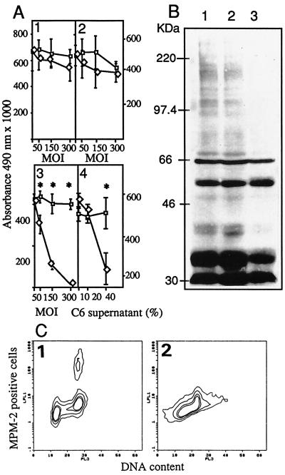
Figure 2.
(A) Inhibition of endothelial cell proliferation. C6 (1), MDA-MB-231 (2), and HMEC-1 (3) were injected with AdK3 (◊) or Ad-CO1 (□). HMEC-1 cells (4) cultured with the supernatant from AdK3- (◊) or AdCO1-infected C6 glioma cells (□). (B) Detection of MPM-2 phosphoepitope in HMEC-1 cells. Mock-infected cells (1), AdCO1-infected cells (2), and AdK3-infected cells (3). (C) MPM-2 epitope were detected in HMEC-1 infected with AdCO1 (1) or AdK3 (2) by indirect immunostaining and DNA content by propidium iodide staining, and quantified by flow cytometry (see Materials and Methods). A Student’s t test was used for statistical analysis.

