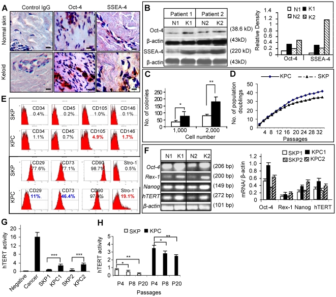
Figure 1. Identification of dermal derived precursor cells from keloid tissues.
A, Frozen sections of keloid tissues and their matched peripheral normal skins were immunostained with specific antibodies for human Oct-4 and SSEA-4. Scale bars, 50 µm. B, Expression of Oct-4 and SSEA-4 in keloids (K1, K2) and matched normal skins (N1, N2) were determined by Western blot analysis and scanning densitometer. C, Colony formation analysis of cells derived from keloids and normal skin (mean±SEM). * P<0.05; ** P<0.01. D, Cell population doubling numbers were determined in single cell colony-derived stem cells from keloids (KPCs) and matched peripheral normal skins (SKPs) by standard 3T3 cell culture protocol (mean±SEM). E, Flow cytometric analysis of cell surface markers of KPCs and SKPs. F, RT-PCR (upper panel) and qPCR (lower panel) analysis of stem cell genes of KPCs (K1, K2) and SKPs (N1, N2). G and H, Analysis of telomerase activity using TeloTAGGG telomerase PCR ELISA kit (mean±SEM). * P<0.05; ** P<0.01; *** P<0.001. Data are representative of at least five independent experiments using specimens obtained from different patient donors with matched normal controls (n = 5).

