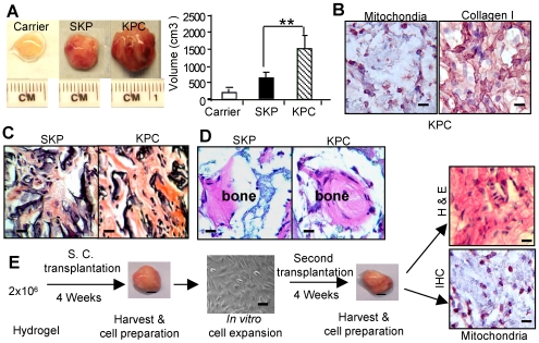
Figure 2. In vivo transplantation of dermal stem cells.
A, Size and volume of transplants generated from SKPs and KPCs using Gelfoam as a carrier for 8 weeks (mean±SEM). ** P<0.01. B, Immunohistological studies of KPC transplant tissues using a specific antibody for human mitochondria (purple color) and type I collagen (brown color), respectively. Scale bars, 50 µm. C, H & E histological stain of transplants. Scale bars, 50 µm. D, In vivo bone regeneration by SKPs or KPCs using hydroxyapatite/tricalcium phosphate (HA/TCP) as carrier. Scale bars, 50 µm. E, Serial transplantation of KPCs. KPCs (2×10⁶) with hydrogel were injected subcutaneously into nude mice. After 4 weeks, the transplanted were harvested and the recovered cells were expanded in vitro and re-transplanted into mice for another 4 weeks. The transplant tissues were harvested for H & E staining or immunohistochemical (IHC) staining with a specific antibody for human mitochondria (purple color). Scale bars, 50 µm. The results are representative of five independent experiments.

