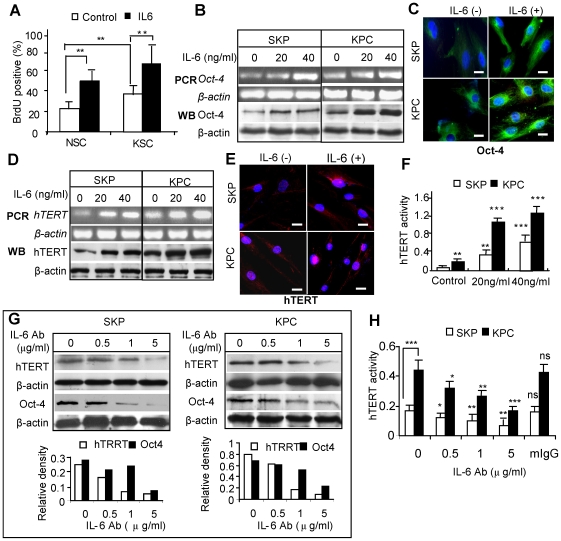
Figure 4. IL-6 increases Oct-4 and telomerase expression in KPCs and SKPs.
A, KPCs and SKPs were cultured in 1% FBS for 24 hours followed by exposure to different concentrations of IL-6, and BrdU incorporation in KPCs and SKPs was determined (mean±SEM). ** P<0.01. B, Expression of Oct-4 mRNA and protein were determined by Western blotting (WB) and RT-PCR, respectively. C, Immunofluorescence studies of Oct-4 expression in SKP and KPC after incubated with 20 ng/ml IL-6 for 24 hours. Scale bars, 20 µm. D, Expression of hTERT mRNA and protein were determined by Western blotting (WB) and RT-PCR, respectively. E, Immunofluorescence studies of hTERT expression in SKP and KPC following incubation with 20 ng/ml IL-6 for 24 hours. Scale bars, 20 µm. F, Telomerase enzyme activity of SKPs and KPCs in response to IL-6 as determined by TeloTAGGG Telomerase PCR ELISA. G and H, Treatment with neutralizing antibody for IL-6 (IL-6Ab) decreased the basal level of hTERT as determined by Western blotting (G) and TeloTAGGG Telomerase PCR ELISA (H). An isotype-matched normal mice IgG (mIgG) was used as negative control (mean±SEM). * P<0.05; ** P<0.01; *** P<0.001; ns, no significance. The results are representative of at least five independent experiments using KPCs and the matched SKPs from different patient donors (n = 5).

