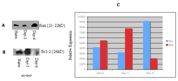
Fig 3. The apoptotic process in T cells from T. gondii infected mice is associated with altered induction of Bcl-2 or Bax.
Cell lysates were prepared from purified T lymphocyte populations as described in the materials and methods. Each lane (A-B) contains lysate of 106 cells. The detection of antigen-antibody complexes was conducted by enhanced chemiluminescence. The level of Bcl-2/ Bax expression intensity is relative to the T cells under the same condition from uninfected mice (given a value of 1) (C). Naïve, T cells from uninfected mice; Day-3, T cells from mice infected for 3 days; Day-6, T cells from 6 days infected mice. The relative intensity of Bcl-2/Bax protein expression was determined by using NIH software. Bax induction was higher in T cells from 6 days infected mice compared to naive or 3 days infected mice. Decreased expression of Bcl-2 was observed in cells from mice infected for 6 days compared to animals infected for 3 days or to naïve mice. Similar results were obtained in three repeated experiments (each group had three mice).

