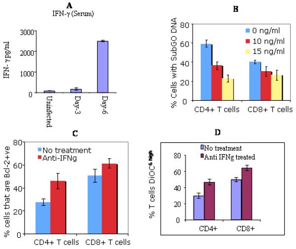
Fig 4. Effects of anti-IFNγ mAb on apoptosis of T cells in T. gondii infected mice.
ELISA measured IFNγ cytokine in the sera from indicated groups of mice (A). Sera from: Naïve, uninfected mice; Day-3, mice infected for 3 days; Day-6, 6 days infected mice. Similar results were obtained in three repeated experiments. Treatment with mAb prevented DNA degradation in a dose depended manner (B). Blockade is more marked in CD4+ T cells (p<0.001) compared to CD8+ T cells (p<0.05) at the concentration 10ng/ml of mAb. Results demonstrated the percent of CD4+ or CD8+ T cells that were Bcl-2 positive (C). Expression of Bcl-2 was significantly increased in CD4+ T cells following treatment with mAb (p<0.001). The restoration of Bcl-2 was less extensive in CD8+ T Cells. treatment increased DiOC6high expression (D). Stained cells (CD4+ and CD8+) were resuspended in 25 nM DiOC6 (see methods and materials) and were analyzed by flow cytometry to assess the loss of mitochondrial transmembrane potential (ΔΨm). Higher percent of CD4+ T cells became DiOC6high after the treatment with anti-IFN . mAb (p<0.001). There was also an increase of DiOC6high expression in CD8+ cells but was relatively less marked than in CD4+ T cells. Data shown in A, B, C, and D are representative of three repeated experiments.

