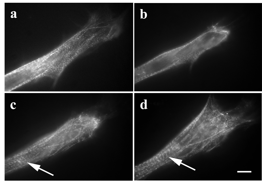
Figure 3.
Recovery of premyofibrils and formation of myofibrils in a myotube after reversal of Lat-A effect. (a) Control myotube transfected with GFP-alpha-actinin. (b) Same myotube after for 25-minute exposure to 5 µM Lat-A. The premyofibrils close to the end of the myotube were lost. (c) and (d) Recovery after the removal of Lat-A: (c) 30 minutes and then (d) 1 hr recovery. The myotube reformed its premyofibrils, and some of them assembled into mature myofibrils (arrows, c, d). Bar = 10 microns.

