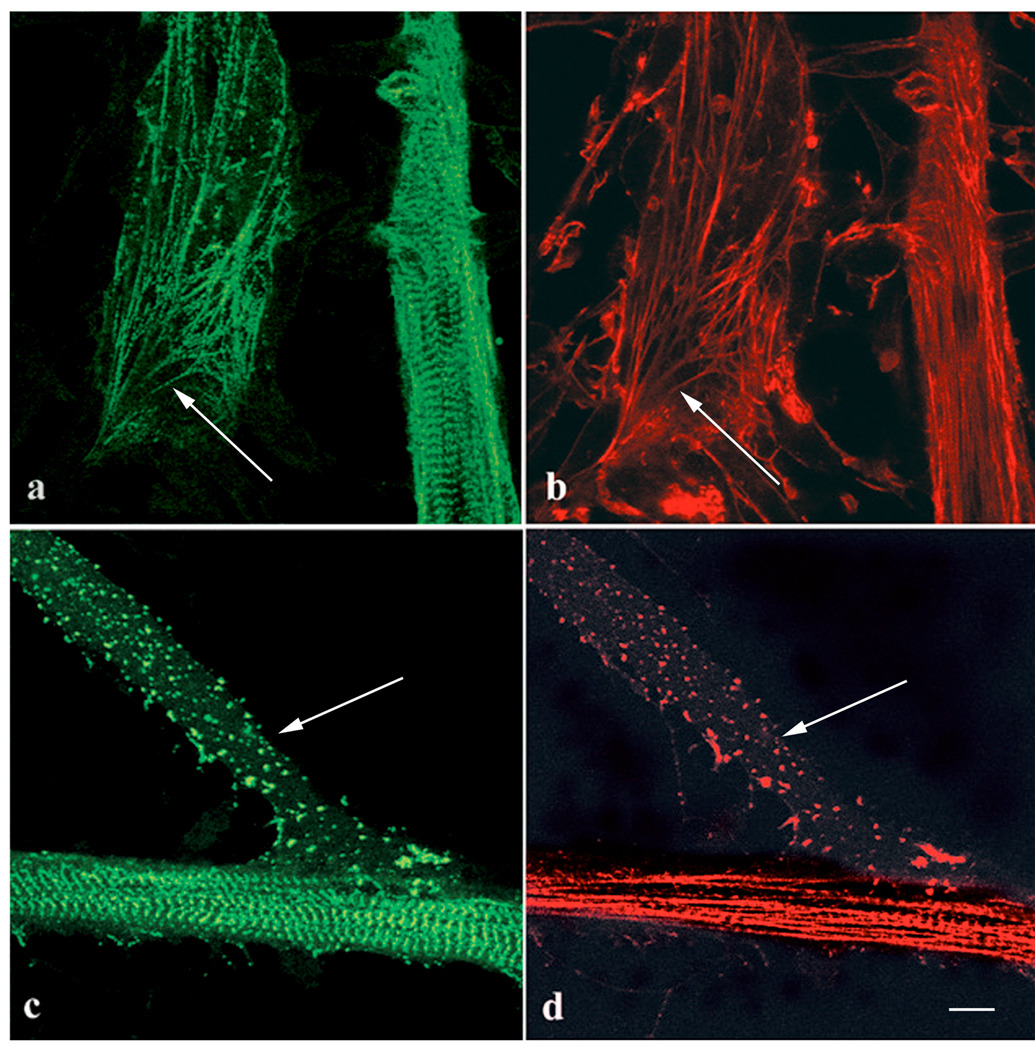
Figure 5.
Control (a, b) and Lat-A treated (c, d) myotubes stained with (a, c) anti-sarcomeric alpha-actinin antibody and (b, d) rhodamine phalloidin. (a, b) At the end of one myotube (arrows), premyofibrils have (a) a banded distribution of alpha-actinin with short sarcomeric spacings and (b) unbanded actin staining. Along the length of the other myotube are mature myofibrils with alpha-actinin in Z-bands and actin filaments that are banded with phalloidin staining. (c, d) Parts of two myotubes treated with 5 µM Lat-A for one hour. Multiple patches of alpha-actinin and actin fill the end of one myotube (arrows) with no sign of premyofibrils. The mature myofibrils in the central region of the second myotube were unaffected by Lat-A. Bar = 10 microns.

