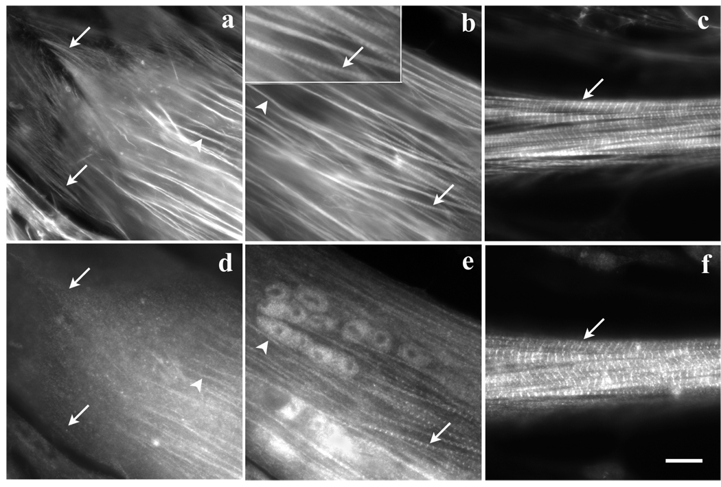
Figure 7.
Three regions of the same myotube stained with (a–c) fluorescently labeled phalloidin and (d–f) anti-CapZ antibodies. (a, d) Actin fibers that extend to the end of the myotube were unstained by the CapZ antibody (arrows in a, d), but some of the more distal fibers (arrowheads) stained with CapZ. (b, e) Fibers in a region of the same myotube near the tip stain with both (b) phalloidin and (e) anti-CapZ antibodies. CapZ was localized in some fibers (arrowheads, b, e) in a continuous pattern and on other fibers (arrows, b, e) it was in a striated pattern with sarcomeric spacings shorter (1.7µm) than the 2 µm sarcomere lengths of mature myofibrils. (c, f) In the rounded central shaft of the same myotube, both (c) phalloidin and (f) anti-CapZ antibodies are in a periodic pattern of 2 µm sarcomeric spacings in the mature myofibrils. The bright band of phalloidin staining is the Z-band where actin filaments overlap. The position of these mature myofibrils corresponds to the region of the myotube where myofibrils were resistant to the effects of Lat-A (see Figure 4 c). Bar = 10 microns.

