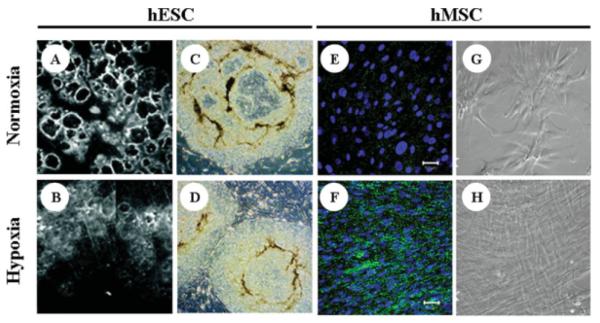
Figure 2. Effect of hypoxia on in vitro stem cell phenotype.
(A and B) Human ESCs (H1) cultured on MEF feeder layers under normoxic (21% O2) and hypoxic (5% O2) conditions. Normoxic cells display extensive regions of differentiation (dark circles surrounded by bright rings) compared with hypoxic cultures. (C and D) Further comparison of H1 hESCs grown at 21% O2 and 3% O2, respectively, showing fewer differentiated (darker, nonuniform) regions under hypoxic conditions. Images adapted from Ref. 50, with permission from National Academy of Sciences. (E–H) Comparisons of bone marrow-derived hMSCs grown under normoxic (21% O2) or hypoxic (2% O2) conditions: (E and F) Significantly higher connexin-43 expression is observed in hypoxic hMSCs grown in monolayer culture for 11 days using expansion medium. (G and H) Phase contrast images demonstrate the maintenance of uniform spindle morphology at late stages of passage under hypoxic conditions. Images adapted from Ref. 61, with permission from Academic Press.

