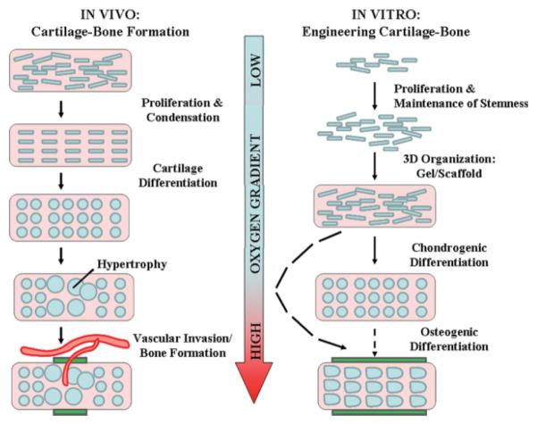
Figure 3. Learning from nature: The influence of oxygen tension during bone-cartilage tissue formation.
In vivo: During development, cartilage forms via the processes of mesenchymal condensation and carefully regulated chondrogenic differentiation in very hypoxic environments. Long bone formation occurs via endochondral ossification where hypertrophic chondrocytes upregulate angiogenic genes triggering vascular invasion (with consequent increases in oxygen tension) followed by new bone formation (green rectangles). Images adapted from Ref. 89, with permission from Nature Publishing Group. In vitro: Hypoxia enables the expansion of stem cells while maintaining their undifferentiated states. Cells are then seeded into 3D organizations to facilitate functional tissue development. Chondrogenic differentiation is enhanced under hypoxic conditions relative to 20% O2. Differentiation into osteogenic lineage and subsequent bone formation (green rectangles) in vitro is optimized at ambient (20%) O2 tensions and inhibited under hypoxic conditions.

