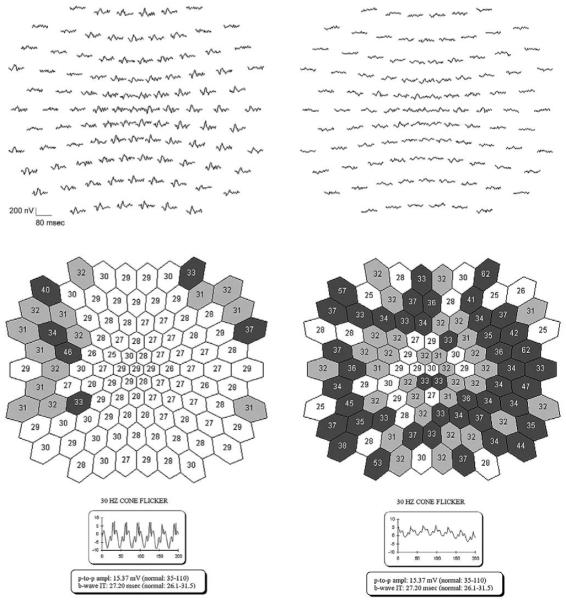
FIGURE 5.
Timing of multifocal electroretinography (mfERG) responses in two patients with RP10 adRP. Representative raw mfERG responses are displayed at top left and right; the full-field responses are presented at the bottom. Middle left and right shows the best fitting values of the timing parameter for the mfERG responses.21 Lighter colors represent less delayed responses; darker colors, more delayed responses. (Left) Patient VI/5, with normal full-field 31 Hz cone response timing, had some delayed responses in the periphery and responses with normal timing in the center of the mfERG. (Right) Patient VI/1, with delayed 31 Hz cone responses, exhibited delayed timing both centrally and peripherally with more substantial delay in the periphery on the mfERG.

