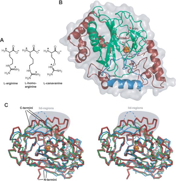
Fig. 3.
A) Chemical structures of the substrates accepted by VioC. B) Overall structure of the substrate complex VioC•L-Arg•tartrate•Fe(II). The β-strands B, G, D, I, and C build the major side of the jelly roll fold and the minor side is built by the β-strands F, E, and H. The flexible lid region is shown in blue, the bound Fe(II) depicted in orange, and the cosubstrate mimic and the substrate are shown in gray. C) A stereo diagram shows the comparison of the ribbon diagram of the VioC•L-Arg•tartrate•Fe(II) complex (red, bold) with the AsnO•hAsn•succinate•Fe(II) complex (green, PDB accession code 2OG7) and with CAS (blue, PDB accession code 1DRY). The position of the iron atom is marked as an orange sphere. The lid regions (VioC: residues F217–P250; AsnO: residues F208–E223; CAS: residues M197–G207 with disordered parts indicated by dashed lines) are highlighted in gray.

