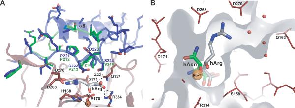
Fig. 6.
Lid-control of substrate binding. A) Comparison of the lid regions of VioC (blue) and AsnO (green). The side chain of residue S224 forms a hydrogen bond to Q137 that coordinates the α-amino group of hArg (distance is indicated in Å). The residues sealing the active site are also specified. B) Superposition of hArg (gray) and hAsn (green) coordination in the active sites of VioC and AsnO. The catalytic iron is shown in orange. Water molecules near the entrance/exit site for substrates and products of VioC are marked in red.

