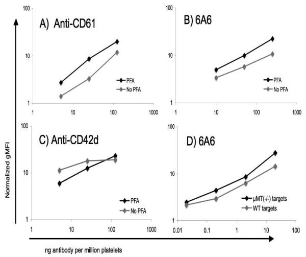
Figure 1. Effect of formalin fixation and μMT(−/−) target platelets on antiplatelet antibody assay sensitivity.
A, B, C) WT platelets were briefly fixed in 1% paraformaldehyde (PFA), or not, and exposed to the indicated amounts of the antibodies shown. Antibody binding was then detected with PE anti-hamster IgG (A, C) or FITC goat-anti-mouse IgG/M (B). D) Paraformaldehyde fixed WT platelets were exposed to the indicated amounts of 6A6 antibody, and antibody binding was detected with FITC goat-anti-mouse IgG/M. All geometric mean fluorescence intensities in the figure were normalized to controls receiving no primary antibody.

