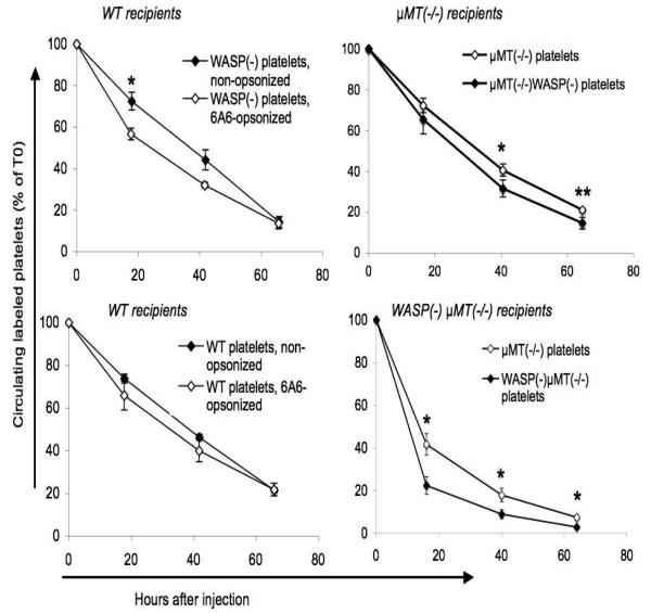
Figure 8. In vivo platelet clearance.
Platelets labeled with fluorescent markers were injected via tail vein. The fraction of labeled platelets was determined at 5 minutes after injection (T0) and at the intervals shown. All error bars are standard errors. Left: Effect of opsonization with 6A6 antibody on platelet clearance. CMFDA-labeled platelets were opsonized with 6A6 antibody and injected into WT recipients. Platelets were identified via log forward vs. log side scatter. N=3 for each type of injected platelet. Right: Effect of μMT(−/−) genotype on platelet clearance. Platelets were labeled with either CMFDA or BMQC. Mixtures of the two genotypes shown in the top or bottom figures were injected into the recipients shown; their clearance was followed simultaneously. Platelets were gated using both log forward vs. log side scatter and a PE-anti-CD41 marker. N=5 recipients in each group. Asterisks indicate p values less than 0.05 (*) or 0.01 (**), (student's two-tailed t-test).

