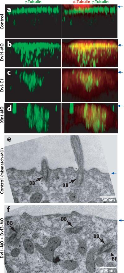
Figure. 3. Dishevelled and Inturned are essential for the apical positioning of basal bodies.
A. Control embryos were stained with anti-γ-tubulin antibody to visualize basal bodies (green). Ciliary microtubules are visualized with anti-α-tubulin antibody (red). Serial confocal images are projected in X-Z plane, with the position of apical membrane indicated by blue arrows at right. B. Failure of apical basal body localization in Dvl1 morphants. C. Failure of apical basal body localization in Dvl-C1 expressing cells. D. Failure of apical basal body localization in Inturned morphants. E. TEM transverse section of control (Dvl1 mismatch MO injected) embryo reveals normal outgrowth of ciliary axonemes from basal bodies docked at the apical cell surface. Cilia can be observed in cross-section projecting above the apical surface. F. Basal bodies fail to dock at the apical membrane and remian in the cytoplasm in Dvl morphants. (BB: basal body, BF: basal foot, m: mitochondria).

