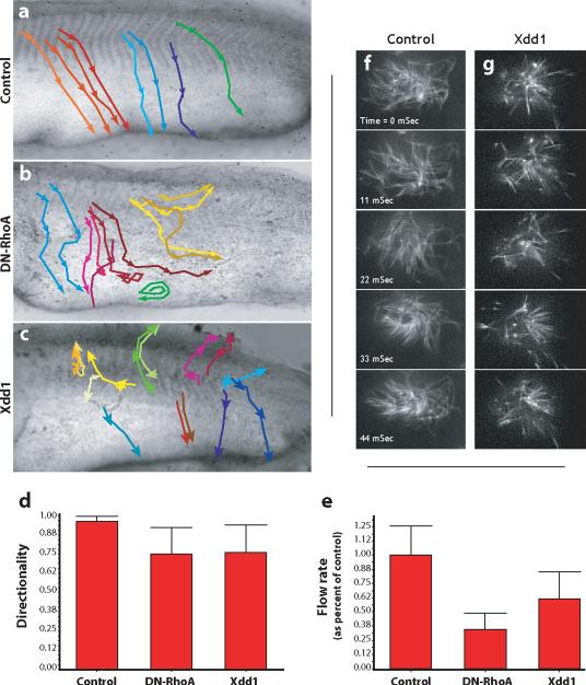
Figure. 8. Dvl and Rho govern directional fluid flow across the ciliated epidermis.
A. Control embryos were grown until St. 26 and latex bead were added to the media to visualize fluid flow over the epidermis. The movement of beads was recorded and representative beads were tracked. Representative bead movements were tracked from the horizontal myoseptum, as indicated by the colored lines. Beads move from dorso-anterior to ventral-posterior uniformly in control embryos. B. DN-RhoA expression severely disrupts directional flow; beads were turning and swirling frequently. C. Xdd1 expression severely disrupts directional flow; beads were turning and swirling frequently. D. The directionality of beads was quantified as the ratio of the linear distance between the first and last point of a track and the total diatance along the track. A straight line would be a value of 1.00. E. DN-RhoA or Xdd1 reduces the rate of bead movement across the epidermis. Finally, defects in polarized flow were evident in that even beads moving in comparatively straight paths moved at a greatly reduced rate across experimental embryos. F. Stills from a high-speed time lapse movie of beating cilia labelled with tau-GFP. G. Stills from a high-speed time lapse movie of beating cilia expressing Xdd1, frequency is comparable to control.

